Thyroid Eye Disease Symptoms, Stages, and Treatment Options
Published on: 18 August 2026
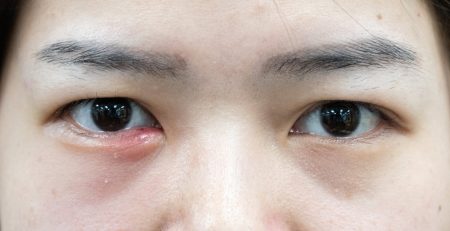
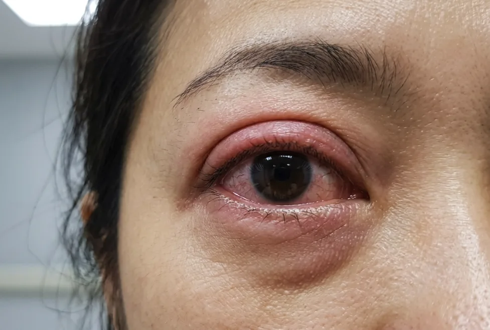
If you have noticed your eyes becoming more prominent, protruding, eyelid retraction, red or uncomfortable, it could be a sign of Thyroid Eye Disease (TED). In Singapore, effective management relies on determining whether the condition is in the active (inflammatory) or inactive (stable) phase. Treatment strategies vary by stage, ranging from conservative symptom relief to targeted medical therapy and phased surgical reconstruction. Timely clinical evaluation by an ophthalmologist or oculoplastic specialist is essential to safeguard vision and restore ocular health.
Key Takeaways
- Thyroid Eye Disease is an autoimmune orbital condition that causes swelling and tissue expansion behind the eyes. It is distinct from systemic thyroid disorders and requires independent ophthalmic management.
- Clinical management depends entirely on the stage of the condition. The Active Phase involves inflammatory changes managed with medical therapies. Structural surgical interventions are planned during the stable, Inactive Phase.
- When clinical rehabilitation is required, procedures follow a standardised medical order: Orbital Decompression → Strabismus Surgery → Eyelid Surgery.
- Because these interventions are performed for functional and reconstructive purposes to safeguard vision, they are typically eligible for MOH Medisave and private insurance claims.
What is Thyroid Eye Disease (Graves’ Ophthalmopathy)?
Thyroid Eye Disease (TED), also known as Graves’ ophthalmopathy or thyroid orbitopathy, is complex autoimmune condition that causes inflammation and swelling behind the eyes, leading to symptoms like bulging (proptosis), double vision, and eyelid retraction. While frequently linked to thyroid gland imbalances, it remains a distinct clinical condition requiring specialised ophthalmic care.
The Autoimmune Root Cause of TED
The root cause of TED occurs when the immune system mistakenly attacks the healthy tissues inside the eye socket. Antibodies bind to cells behind the eye, triggering inflammation and causing tissues to absorb excess water. Because the bony eye socket cannot expand, the swollen muscles and fat push the eyeball forward and restrict its movement, leading to the physical symptoms of the condition.
The Relationship Between TED and Systemic Thyroid Levels
While TED is a separate condition from systemic thyroid issues, it is closely tied to Graves’ disease. Studies show that roughly 80% to 90% of patients with orbital inflammation also have an overactive thyroid. However, TED can still affect people with an underactive or even normal thyroid. Because the two conditions run on different pathological tracks, simply normalising your blood thyroid levels will not automatically cure or stop the active changes happening behind your eyes.
Key Risk Factors and Patient Demographics in Singapore
The risk of developing this condition is influenced by several clinical and environmental factors:
- Tobacco Use: Smoking is a modifiable risk factor, multiplying the likelihood of severe disease progression by several times.
- Gender: The condition is more common in females, although males are more likely to develop severe, sight-threatening forms.
- Age of Onset: While Western populations typically present in their late 40s or 50s, research shows that local asian patients often present younger, with a mean onset age of approximately 40 years.
- Poor Thyroid Control: Unstable thyroid hormone levels can increase the severity of active orbital inflammation.
Recognising TED Symptoms: How It Affects Asian Eyes Differently
Early identification of symptoms helps prevent permanent tissue changes. Anatomical variations in Asian eyelid structures can affect how physical changes present clinically.
Early Warning Signs and Initial Symptoms
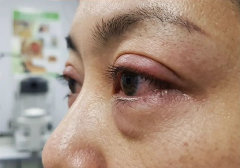
The early stages of the disease are often mistaken for common eye allergies or chronic dry eye syndrome. These early warning signs are usually more prominent in the morning due to fluid accumulation overnight. The most common early symptoms include:
- A gritty, sandy sensation under the eyelids
- Excessive tearing and watery eyes
- Unusual sensitivity to bright light (photophobia)
- Puffiness or redness of the upper and lower eyelids
- A dull, deep ache behind the eyes, especially when looking in different directions
Proptosis (Bulging Eyes) and Eyelid Retraction
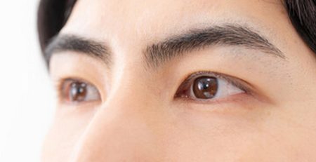
As tissues behind the eye continue to swell, they push the eyeball forward, a condition known as proptosis or exophthalmos. This forward movement is often paired with eyelid retraction, where the upper eyelid sits abnormally high or the lower eyelid sits too low. This retraction exposes the white of the eye (the sclera) above and below the cornea, creating a constant startled or staring look. This exposure prevents proper blinking, leaving the cornea vulnerable to dry spots and painful ulcerations.
Clinical Differences: Asian vs. Caucasian Presentation
Clinical data from Southeast Asian and East Asian cohorts reveal distinct differences in how Thyroid Eye Disease manifests compared to Caucasian patients.
| Clinical Marker | Asian Patient Presentation | Caucasian Patient Presentation |
| Average Bulging (Proptosis) | Lower baseline exophthalmometry readings (mean ~18.8mm to to 19.0 mm) | Substantially higher baseline bulging measurements (20.1–22.3 mm) |
| Eyelid Alignment Issues | High incidence of acquired epiblepharon (lashes turning inward) | Uncommon; primary presentation is simple eyelid retraction |
| Corneal Complications | Higher rate of corneal erosions (nearly 30%) due to lash rubbing | Lower incidence of primary lash-related corneal friction |
| Orbital Space Dynamics | Narrower orbital apex, increasing risk of early nerve compression | Wider orbital apex, allowing more tissue expansion before compression |
Diagnosing and Grading Thyroid Eye Disease
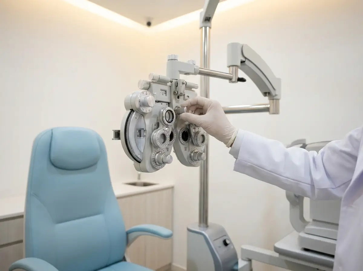
Accurately diagnosing and grading Thyroid Eye Disease requires a thorough clinical assessment. Specialists evaluate the active inflammation and measure the physical changes to prevent potential vision loss.
Clinical Eye Examinations and the Clinical Activity Score (CAS)
A detailed evaluation begins with measuring visual acuity, eye pressure, and colour vision. To determine the phase of the disease, specialists use the Clinical Activity Score (CAS), which is a seven-point diagnostic system. One point is awarded for each of the following active inflammatory signs:
- Spontaneous pain behind the eye.
- Pain on upward or downward gaze.
- Eyelid redness.
- Eyelid swelling (edema).
- Conjunctival redness.
- Conjunctival swelling (chemosis).
- Inflammation of the caruncle.
A CAS score of 3 or higher indicates active disease, which requires immediate anti-inflammatory medical treatment.
Orbital Imaging and Optic Nerve Assessment
High-resolution Computed Tomography (CT) or Magnetic Resonance Imaging (MRI) scans provide detailed cross-sectional views of the eye socket. MRI is effective for detecting active muscle edema, while CT evaluates bony structures prior to surgical planning.
Scans also help detect Dysthyroid Optic Neuropathy (DON), a severe condition where swollen extraocular muscles compress the optic nerve at the back of the socket. Specialists evaluate visual field sensitivity, pupillary responses, and colour perception to detect optic nerve stress early.
Diagnosing Optic Nerve Compression (Optic Neuropathy)
Dysthyroid Optic Neuropathy (DON) is a rare but severe complication where swollen eye muscles compress the optic nerve, threatening permanent vision loss. To catch this early, specialists perform visual field examinations, test pupillary responses, and evaluate colour vision. Any decline in colour vibrancy or loss of peripheral vision requires immediate clinical intervention to relieve the pressure on the nerve.
The Two Phases of TED: Active vs. Inactive Stages
TED follows a clinical progression known as Rundle’s Curve. This curve consists of an initial active inflammatory phase followed by a stable, inactive phase.
| Phase 1: Active Inflammatory | Peak Severity | Phase 2: Inactive / Quiescent |
| Active inflammation & swellingHigh Clinical Activity Score (CAS)Managed with anti-inflammatory medical therapy | Maximum point of orbital tissue expansion and symptoms | Fibrotic scarring & stable changesLow Clinical Activity Score (CAS)Safe window for restorative surgeries |
Timeline and Management of Triggers
The active phase typically lasts between 18 and 24 months. During this period, symptoms may fluctuate. Reconstructive surgeries are postponed during the active phase, as shifting inflammation makes surgical predictions unreliable. Key preventive measures during this period include:
- Complete Smoking Cessation: Stopping all tobacco use reduces the risk of prolonged active phases.
- Thyroid Stabilisation: Working closely with an endocrinologist helps maintain stable blood hormone levels.
- Cautious Radioiodine Use: Radioactive iodine (RAI) therapy for hyperthyroidism can trigger orbital flares unless accompanied by prophylactic corticosteroid coverage as directed by a doctor.
Non-Surgical Management During the Active Phase
During the active stage of TED, clinical goals focus on relieving daily discomfort, protect the ocular surface, and suppress active immune-mediated inflammation.
Conservative Relief and Daily Care
- Ocular Lubrication: Preservative-free artificial tears during the day and lubricating ointments at night protect the cornea.
- Head Elevation: Sleeping with the head elevated reduces nocturnal fluid retention and morning swelling.
- Prism Lenses: Temporary Fresnel prisms attached to spectacles can align diplopia (double vision) for functional daily vision.
Medical and Biologic Therapies
For moderate-to-severe active disease, specialists may prescribe targeted medical interventions:
- Intravenous Corticosteroids: Pulses of intravenous methylprednisolone are commonly used to reduce active orbital inflammation and relieve pressure on the optic nerve.
- Targeted Biologic Agents: Teprotumumab (Tepezza) is a targeted monoclonal antibody approved for use in Singapore. By specifically blocking the insulin-like growth factor-1 receptor (IGF-1R) involved in the underlying disease mechanism, clinical studies show it effectively reduces active inflammation, double vision, and eye bulging (proptosis) in suitable active cases.
- Selenium Supplementation: Evidence suggests daily selenium supplementation for six months may help improve quality of life and mild symptoms in select patients.
Surgical Rehabilitation During the Inactive Phase
Once inflammation subsides and the disease remains stable (typically verified by unchanged clinical measurements for at least six consecutive months), surgical reconstruction can be safely evaluated.
3-Step Reconstructive Sequence
Surgical rehabilitation follows a strict order. Because each stage alters the physical alignment of surrounding tissues, altering this order can compromise final outcomes.
- Orbital Decompression: The surgeon removes targeted portions of the bony socket walls or excess orbital fat. This expands the available space, allowing the eyeball to settle back into a natural position and relieving pressure on the optic nerve.
- Strabismus Surgery: Active inflammation can leave eye muscles scarred or tight. After decompression, strabismus surgery realigns restricted extraocular muscles to correct double vision and restore single binocular vision.
- Oculoplastic Eyelid Surgery: The final step addresses persistent eyelid retraction or asymmetry. Oculoplastic techniques reposition retracted eyelids to protect the cornea and restore symmetry.
Medisave, Insurance, and Treatment Costs in Singapore
In Singapore, surgical treatments for stable Thyroid Eye Disease are classified as functional reconstructive procedures rather than cosmetic enhancements.
Because procedures such as orbital decompression, strabismus correction, and eyelid retraction repair serve to protect vision and rehabilitate functional impairment, eligible portions may be claimed under the Ministry of Health (MOH) Medisave scheme and private Integrated Shield Plans, subject to individual policy limits and doctor assessment.
Red Flag Symptoms Requiring Urgent Assessment
Certain clinical changes require prompt evaluation by an eye specialist or emergency care provider. Contact a medical professional immediately if you experience:
- A sudden or progressive decline in your vision
- Changes in colour perception, such as colours appearing washed out or grey
- An inability to close your eyelids completely, leading to severe pain
- Rapidly worsening bulging of one or both eyes
- Systemic signs of a thyroid storm, including a rapid heart rate, high fever, or severe agitation
Take Control of Your Eye Health
Thyroid Eye Disease is a progressive condition, but timely clinical evaluation during the active phase is key to preventing long-term structural changes and safeguarding your sight. Whether you are experiencing early symptoms like dry eyes and puffiness, or are seeking surgical rehabilitation for stable proptosis, early specialist involvement ensures your care is tailored to your exact phase of disease. Schedule a clinical eye assessment to evaluate your condition and discuss your options.
Take the Next Step
Early detection and treatment are crucial to safeguarding your vision from the irreversible damage.
Book the Comprehensive $88 Cataract & Glaucoma Screening.
Book a Cataract & Glaucoma Screening for $88
Don’t wait for symptoms to appear before taking action. Early detection is key to preserving your vision.
At Dr Roy Tan’s clinic, we offer a comprehensive Cataract & Glaucoma Screening for just $88. This screening is designed to assess your eye health thoroughly, identify any early indicators of glaucoma or cataracts, and discuss your personalised options if either condition is detected.