Proptosis (Bulging Eyes): Causes, Symptoms & Treatment in Singapore
Published on: 09 September 2026
Proptosis (bulging eyes) occurs when one or both eyes protrude forward out of the eye socket. Most commonly caused by Thyroid Eye Disease (TED), it can also stem from orbital infections, vascular anomalies, or tumours. Treatment ranges from targeted medical therapies to specialised 3-stage surgical rehabilitation. Early diagnosis by an oculoplastic specialist is essential to protect vision and prevent corneal damage.
Noticing that one or both of your eyes appear unusually prominent or pushed forward can be distressing. Proptosis is a medical condition where the eyeball protrudes forward from its protective bony socket. Prompt evaluation by a specialist ensures accurate diagnosis, preserves visual function, and achieves optimal cosmetic balance.
What is Proptosis (Bulging Eyes)?
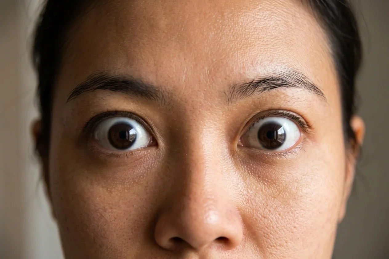
Proptosis means one or both of your eyes bulge forward more than normal. It happens when something inside the eye socket, such as swelling, inflammation, or a tissue growth, pushes the eyeball outward. Identifying the underlying cause early helps protect your vision and prevents long-term discomfort.
Definition & Difference Between Proptosis vs. Exophthalmos
Doctors often use “proptosis” and “exophthalmos” interchangeably to describe bulging eyes, but there is a slight distinction in the medical field. Proptosis is the broader term for an eyeball pushing forward due to issues like swelling, injury, or tissue growth. On the other hand, doctors usually reserve “exophthalmos” for eye bulging caused specifically by thyroid conditions, such as Graves’ disease.
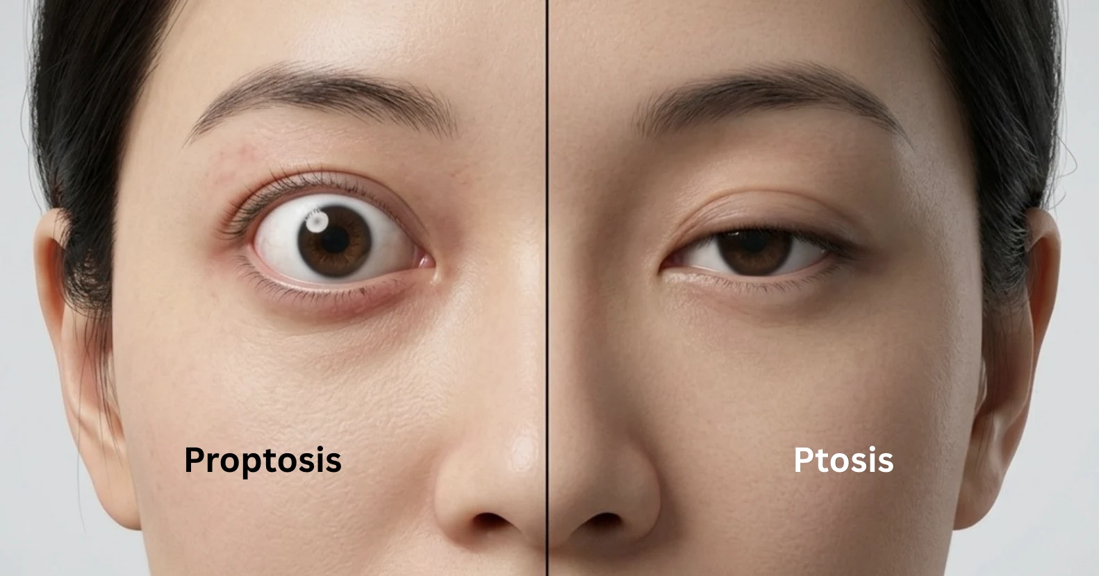
Key Differences: Proptosis vs. Ptosis (Drooping Eyelid)
Proptosis and ptosis are distinct ocular conditions that affect different structures of the eye assembly:
| Feature | Proptosis | Ptosis |
| Primary Anatomical Affected Area | Eyeball (Globe) | Upper Eyelid |
| Physical Appearance | Eyeball protrudes or bulges forward | Upper eyelid droops over the eye |
| Underlying Mechanism | Increased volume or pressure within the orbit | Weakness of the levator muscle or nerve impairment |
| Functional Impact | Exposure keratitis, double vision, orbital pressure | Blocked upper visual field, asymmetrical appearance |
Unilateral vs. Bilateral Proptosis: Why Eye Involvement Matters
The presentation of proptosis in one eye (unilateral) versus both eyes (bilateral) provides critical diagnostic clues regarding the underlying cause:
- Unilateral Proptosis: Involves only one eye. It frequently points towards localised orbital issues such as primary orbital tumours, bacterial cellulitis, vascular malformations, or localised trauma.
- Bilateral Proptosis: Affects both eyes simultaneously. It strongly suggests systemic health conditions, most notably autoimmune thyroid disorders like Graves’ ophthalmopathy.
What Causes Proptosis in Adults and Children?
Proptosis develops when the space within the rigid, bony eye socket becomes compromised by swelling, inflammation, mass lesions, or fluid accumulation. Identifying the root cause guides the treatment approach.
Thyroid Eye Disease (TED) & Graves’ Ophthalmopathy (Most Common Cause)
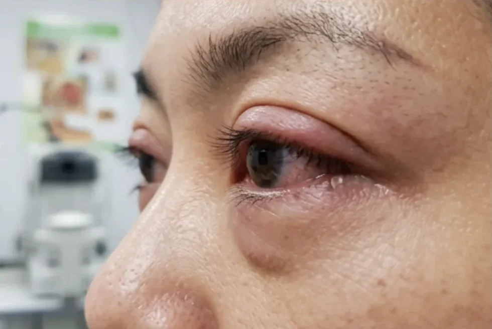
Thyroid Eye Disease (TED) stands as the primary cause of proptosis in adults. This autoimmune condition causes the body’s immune system to attack the soft tissues, fat, and extraocular muscles behind the eye. Inflammation leads to tissue swelling and expansion within the fixed bony orbit, forcing the eyeball forward. Patients may experience TED even when their systemic thyroid hormone levels appear stable under medical control.
Acute Orbital Infections & Cellulitis
Orbital cellulitis is a severe, deep-tissue infection of the fat and muscles surrounding the eye, often arising from spreading sinus infections. The rapid accumulation of pus and inflammatory fluid generates intense orbital pressure, pushing the eye outward. This condition represents a serious medical emergency that requires swift hospital admission and intravenous antibiotic therapy to prevent permanent vision loss or central nervous system complications.
Orbital Tumours, Vascular Malformations, and Trauma
Space-occupying lesions within the orbit directly push the eyeball forward as they expand. These include:
- Benign and Malignant Tumours: Hemangiomas, schwannomas, or lymphomas growing behind the globe.
- Vascular Anomalies: Carotid-cavernous fistulas or orbital varices that alter blood flow dynamics, increasing orbital pressure.
- Orbital Trauma: Fractures and blunt force injuries that cause retrobulbar haemorrhages (bleeding behind the eye), leading to sudden, painful protrusion.
Paediatric vs. Adult Proptosis Causes
Proptosis in paediatric patients demands immediate, specialised assessment. While adult cases stem largely from autoimmune thyroid disorders, paediatric cases often arise from acute orbital infections, congenital vascular lesions, or developmental abnormalities. Rare paediatric orbital malignancies, such as rhabdomyosarcoma or neuroblastoma metastasis, must also be promptly evaluated and ruled out.
What Are the Key Symptoms of Bulging Eyes?
Symptoms accompanying proptosis range from mild surface irritation to severe visual disruption, depending on the speed of onset and degree of protrusion.
Surface & Visual Changes (Dry Eye, Double Vision, Eye Redness)
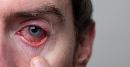
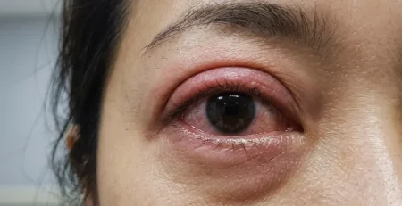
As the eye is pushed forward, the eyelids struggle to cover the front surface fully during normal blinking. This incomplete closure exposes the delicate cornea to surrounding air, triggering chronic dry eye symptoms, constant tearing, and persistent ocular redness. Additionally, when inflammatory swelling affects the extraocular muscles responsible for eye movement, patients frequently experience double vision (diplopia), making everyday activities like reading or driving difficult.
Structural Changes (Eyelid Retraction & Inability to Close Eyes)
Structural adjustments around the eye socket alter facial symmetry and ocular safety:
- Eyelid Retraction: The upper or lower eyelids pull back tightly, exposing white sclera above or below the iris.
- Lagophthalmos: The inability to close the eyelids completely, especially during sleep.
- Corneal Exposure: Prolonged exposure increases the risk of corneal abrasions, persistent epithelial defects, and corneal ulcers.
Red Flag Emergency Symptoms: When to Seek Immediate Medical Attention
Certain clinical warning signs require emergency medical evaluation to protect vision:
- Sudden, rapid onset or worsening of eye protrusion
- Severe, throbbing eye socket pain or headache
- Rapid drop in visual acuity or dimming of vision
- Loss of colour perception (especially red saturation)
- New or worsening double vision accompanied by fever
- Inability to move the eye in any direction
Diagnostic Evaluation: How Oculoplastic Specialists Diagnose Proptosis
Accurate diagnosis requires detailed clinical assessment combined with advanced imaging techniques to pinpoint the precise location and cause of orbital pressure.
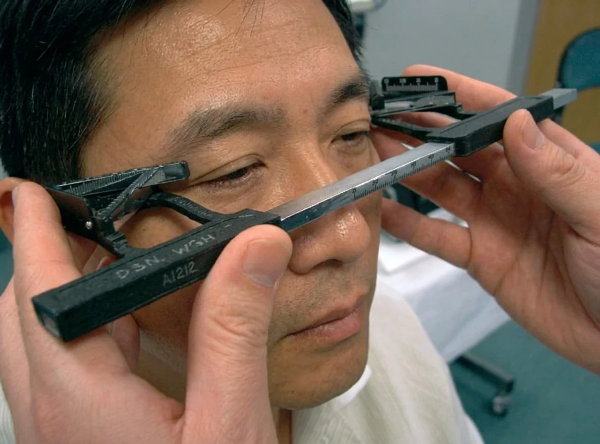
Comprehensive Oculoplastic & Orbital Examination (Hertel Exophthalmometry)
An oculoplastic specialist evaluates eyelid position, extraocular muscle mobility, pupil responses, and corneal integrity. A key clinical tool used during this evaluation is the Hertel exophthalmometer. This specialised instrument measures the exact degree of forward displacement of the cornea relative to the outer edge of the bony orbital rim, establishing a reliable quantitative baseline to monitor disease progression or treatment response over time.
Advanced Imaging: CT Scans & MRI of the Orbit
High-resolution diagnostic imaging provides detailed visualisation of orbital anatomy behind the eye:
- Computed Tomography (CT) Scans: Excellent for evaluating the bony boundaries of the orbit, assessing bone erosion, and detecting orbital fractures or calcifications.
- Magnetic Resonance Imaging (MRI): Superior for evaluating soft tissue detail, such as extraocular muscle enlargement, optic nerve compression, retrobulbar fat volume, and orbital mass characteristics.
Systemic Blood Tests & Thyroid Function Tests
Blood tests help identify the underlying root causes of your condition that scans cannot see. They primarily check your thyroid hormone levels, including TSH (thyroid-stimulating hormone, a brain hormone that tells your thyroid to make and release energy-controlling hormones), free T3 and free T4 (active hormones that control your body’s energy), and screen for specific autoimmune antibodies, such as TSI (thyroid-stimulating immunoglobulin, an antibody that forces the thyroid to overproduce hormones) and TRAb (TSH receptor antibodies, immune proteins that attach to the thyroid and disrupt its normal signalling). Depending on your symptoms, your doctor may also test for general body inflammation or active infections.
Proptosis Treatment Options in Singapore
Bulging eyes can be managed through both non-surgical and surgical options depending on the underlying cause, severity, and impact on your vision.
Non-Surgical Treatments & Medical Management (Steroids, Biologics)
During active inflammatory phases, non-surgical therapies help control disease progression:
- Intravenous Steroid Therapy: High-dose corticosteroid infusions rapidly suppress active orbital inflammation and decrease soft tissue swelling.
- Targeted Biologics: Modern immunomodulatory medications directly target inflammatory pathways responsible for autoimmune tissue remodelling. Breakthrough targeted therapies like Tepezza (teprotumumab), an IGF-1 receptor inhibitor designed specifically for Thyroid Eye Disease, block the underlying signaling that drives orbital inflammation, muscle swelling, and tissue expansion.
- Lubricating Eye Drops and Ointments: High-viscosity artificial tears protect the exposed corneal surface from severe dry eye and ulceration.
- Prism Lenses: Specialised spectacle lenses help realign double vision caused by swollen extraocular muscles.
Surgical Rehabilitation: The 3-Stage Treatment Approach
When conservative treatments prove insufficient, or during the stable phase of condition management, specialised surgical procedures help restore function and appearance.
Stage 1: Orbital Decompression Surgery
This primary procedure creates extra space within the tight eye socket. The oculoplastic surgeon carefully removes select portions of the bony orbital walls, removes excess orbital fat, or combines both techniques. This allows the bulged eyeball to settle back into a natural position within the socket, immediately relieving pressure on the optic nerve and reducing corneal exposure.
Stage 2: Strabismus (Eye Muscle) Surgery
Once the eye returns to a safer position within the socket, persistent double vision may remain due to scarred or tight extraocular muscles. Strabismus surgery adjusts, repositions, or loosens these eye muscles to realign both eyes, restoring comfortable single binocular vision when looking straight ahead and reading.
Stage 3: Eyelid Margin & Retraction Surgery
The final stage addresses remaining eyelid abnormalities. Surgical repositioning relaxes retracted upper or lower eyelids, improving the eye’s natural closure mechanism (lagophthalmos), protecting the cornea, and refining cosmetic symmetry.
Treatment Cost in Singapore
Proptosis and Thyroid Eye Disease (TED) treatment costs vary based on clinical complexity, diagnostic imaging requirements, facility fees, and the overall scope of surgical intervention.
- Initial Specialist Consultation: Specialist evaluations in private practice typically range between $88 and $450, depending on consultation duration and preliminary testing.
- Diagnostic Imaging (CT/MRI Orbit): Specialised orbital imaging ranges from $1,000 to $2,500, depending on soft tissue detail, contrast requirements, and facility settings.
- Orbital Decompression Surgery: Complex surgical procedures vary widely depending on whether single-wall, multi-wall, or fat-only decompression is required. Overall costs generally range from $8,000 to $25,000 per eye, which includes surgeon fees, general anesthesia, and day surgery or inpatient facility expenses.
Insurance, Integrated Shield Plans, and MediSave Coverage for Proptosis
Proptosis management driven by underlying conditions such as Thyroid Eye Disease, orbital cellulitis, or space-occupying tumours is classified as a medically necessary reconstructive procedure rather than a cosmetic enhancement.
Singaporeans and Permanent Residents can utilise MediSave savings for eligible surgical procedures and inpatient stays, subject to prevailing Ministry of Health (MOH) withdrawal limits. Furthermore, private Integrated Shield Plans (IPs) and corporate health insurance policies generally cover hospitalisation, diagnostic scans, and reconstructive surgical procedures for therapeutic indications.
Expected Recovery Timeline and Long-Term Prognosis
Recovery timelines depend on the specific surgical interventions performed:
- Initial Post-Operative Phase (Weeks 1–2): Mild bruising, swelling, and temporary discomfort around the eye socket are expected, requiring prescribed eye drops and rest.
- Functional Stabilisation (Weeks 4–8): Most swelling settles, allowing for initial evaluation of eyeball positioning and muscle alignment.
- Final Rehabilitation (Months 3–6): Staged surgeries (such as muscle or eyelid adjustments) are scheduled sequentially, leading to long-term structural stability, improved visual comfort, and restored quality of life.
Why Choose Dr Roy Tan at Cornerstone Eye Centre
Selecting a dedicated specialist ensures comprehensive care throughout your treatment journey. Dr Roy Tan brings extensive sub-specialty experience in oculoplastic, orbital, and reconstructive eye surgery. Situated conveniently at Novena and Mount Alvernia, Cornerstone Eye Centre offers tailored diagnostic protocols, modern surgical facilities, and patient-centred clinical care focused on safeguarding both vision and structural aesthetics.
Book a consultation to discuss your vision and treatment options.
Schedule an Eye Assessment in Singapore
If persistent eye fatigue or dryness is affecting your daily comfort and work productivity, professional guidance can help identify the root cause. Schedule an appointment to evaluate your tear film stability, check for uncorrected refractive errors, and discuss personalised relief options.
Take the Next Step
Early detection and treatment are crucial to safeguarding your vision from the irreversible damage.
Book the Comprehensive $88 Cataract & Glaucoma Screening.
Book a Cataract & Glaucoma Screening for $88
Don’t wait for symptoms to appear before taking action. Early detection is key to preserving your vision.
At Dr Roy Tan’s clinic, we offer a comprehensive Cataract & Glaucoma Screening for just $88. This screening is designed to assess your eye health thoroughly, identify any early indicators of glaucoma or cataracts, and discuss your personalised options if either condition is detected.